Piezo vs Cavitron. Which ultrasonic vibrations have a greater damaging effect to a tooth surface?
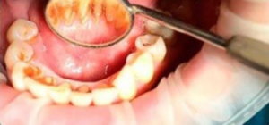
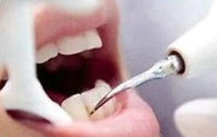
In recent years, there has been an increase in the number of aesthetic restorations of both anterior and posterior groups of teeth with various restorative materials. One of the components of maintaining the integrity of previously performed restorations is professional oral hygiene using both manual and mechanical tools. Most of the dental hygienists use ultrasonic devices in their practice for scaling the teeth. Dental market offers mainly 2 types of ultrasonic devices based on piezoelectric and magnetostrictive types of ultrasonic vibrations.
The use of ultrasound provides several advantages: the speed and ease of manipulation, irrigation of the wound surface with medications.
But there is a downside. Kim SY, Kang MK, and colleagues investigated the effects of employing an ultrasonic instrument on a range of enamel abnormalities. Ultrasonic scaling can severely damage surfaces including as fractured enamel, early caries, and resin restorations.
Another group of researchers—A. I. Grudyanov, K. E. Moskalev and A. V. Sizikov noted that the use of standard nozzles of the Piezo ultrasonic device at medium power for processing the tooth surface can lead to cracking of the surface of the gingival part of composite restorations.
What effect does the ultrasonic tip directly have on the restoration and which ultrasonic vibrations (piezoelectric or magnetostrictive) have a greater damaging effect? They examined the surfaces of various repair structures following piezoelectric and magnetostrictive ultrasonic vibrations to find the answer to the question.
A composite filling, a ceramic veneer, a metal-ceramic crown and an all-ceramic crown were taken as samples for the study. 56 surfaces of composite fillings, 72 surfaces of metal-ceramic crowns, of which 36 were buccal surfaces, 36 were lingual surfaces, and 10 surfaces of ceramic veneers were subjected to the study. All studied samples were treated with two types of ultrasonic vibrations, namely: piezoelectric, generated by the Piezon-Master 400 device, and magnetostrictive, generated by the Cavitron Select device using standard ultrasonic tips to remove supragingival dental plaque. As objects of comparison, samples of untreated surfaces (tooth enamel, composite filling, all-ceramic crown) were taken.
The study was conducted on the surface of direct and indirect restorations in the gingival area, at 1-2 mm from the edge of the restoration. The quality of the marginal fit was not included in this study. The studied samples were processed within 2 minutes. To compare the effects of ultrasonic vibrations on restorative structures, 10 samples of metal-ceramic crowns processed on the buccal surfaces for 1 minute were chosen.
The experimental stage of the study consisted of scanning electron microscopy (SEM) and laser non-contact profilometry (LBP). SEM examination was carried out using a magnification of 500 and 1000 times and consisted in studying the microrelief of surfaces and identifying damage. Laser non-contact profilometric study (LBP) was carried out using a laser profilograph, which today is a modern means of measuring the geometric parameters of the surface.
The profilometric study data were studied in terms of Ra and Rz:
- Ra – gives an idea of the overall uniformly distributed roughness.
- Rz – characterizes individual large defects.
Results of research.
Scanning electron microscopic examination (SEM)
When visualizing, evaluating and studying SEM of various surfaces (composite fillings, veneers, metal-ceramic crowns) after their treatment with the ultrasonic nozzle of the Piezo device and the Caviton device. Significant differences in the relief of the studied surfaces were found.
SEM of the surface of a carefully polished composite filling was used as a control. The electron diffraction pattern revealed a homogenous surface that is smooth and uniform, with no evident large disruptions in the relief.
Composite filling.
After processing the surface of the composite filling with a Piezo device, the resulting defects are visible on its surface in the form of shallow erosions alternating with smooth areas. While using the Cavitron, a fairly smooth surface is formed with areas of slight roughness.
All-ceramic crown.
On the electron diffraction pattern of the all-ceramic crown surface, chosen as a control, except for small artifacts, no changes in the relief were noted.
When evaluating the electron diffraction patterns of the surfaces of ceramic veneers treated with two ultrasonic devices, significant differences are visualized. So, after processing with the Piezo device, deep multiple cavities of a polygonal shape remain on the surface of the veneer. Also noted are small and few artifacts, which are the remnants of deposition. The Cavitron device demonstrated its non-aggressive nature, resulting in a relatively smooth veneer surface with few defects.
Metal-ceramic crowns.
The same trend was noted in the processing of metal-ceramic crowns. On the electron diffraction pattern, processed from the lingual side with Piezo device, large defects of various sizes and shapes were revealed. The results of the study processed by the Cavitron, recorded minor damage to the ceramics.
Comparing the data obtained, we can say that both ultrasonic devices have demonstrated their destructive power. But on the surface of various restoration structures treated with the piezo ultrasonic device, compared to the impact of the Cavitron, there are more damages (potholes, deformations), and they are larger. This was especially evident when processing the lingual surface of metal-ceramic crowns, which were tested for 2 minutes.
Laser non-contact profilometric study (LBP).
The same samples were tested. The results of profilometry, expressed in quantitative measurements of the microrelief of the surfaces of various restoration structures (Ra and Rz values) after their treatment with nozzles with various sources of ultrasonic vibrations, are shown below on the table.
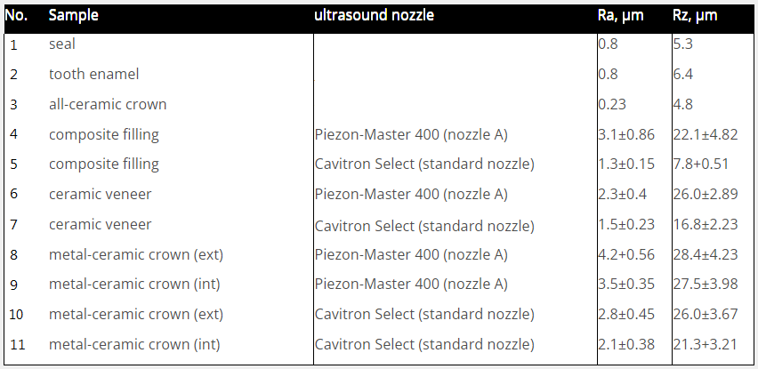
The Ra and Rz values (p < 0.05) when treating the surfaces of a composite filling, ceramic veneer, ceramic-metal crown with Piezo device (sample No. 4, 6, 8, 9) are higher than the Ra and Rz values ( p<0.05) when treating the surfaces of the same samples, but with Cavitron (sample No. 5, 7, 10, 11).
Thus, according to profilometry data, a greater number of defects were obtained during the processing of various surfaces with an ultrasonic nozzle of the Piezo device.
Conclusions
Having the opportunity to study this scientific research, we understand that the procedure of professional teeth cleaning is not as safe as it seems at first glance. We can easily harm the patient. It should be noted that according to previous studies, with a roughness of more than 0.2 microns, favorable conditions are created for the primary adhesion of microorganisms. With the naked eye, we do not see the formed microscopic defects. After some time, the patient pays attention to such changes as the appearance of pigmentation on the restored surface, staining of the edge of the restoration, the rapid formation of plaque.
These complaints from the patient should be an important signal for analysis and correction of subsequent actions by the specialist.
With this in mind, we are obliged to professionally approach these manipulations and practically follow recommendations:
1. When planning the performance of professional oral hygiene for patients with periodontal tissue diseases, a specialist (dentist, periodontist, hygienist) should make a choice of ultrasound devices, considering the volume and amount of soft plaque and calculus, as well as the presence of restoration structures in the oral cavity.
2. When conducting professional oral hygiene in patients with restorations, it is advisable to use ultrasonic devices that generate ultrasound in a magnetostrictive way to remove hard dental deposits.
3. The time of ultrasonic exposure in total should not exceed 2 minutes per surface. The recommended power of the ultrasonic handpiece is medium or low.
4. After using ultrasound, it is recommended to carefully polish the surfaces with certified systems for polishing restorations to prevent the deposition of soft plaque.
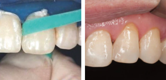
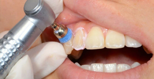
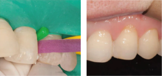
Both types of ultrasonic devices damage teeth surface. Piezo damages more than Cavitron. Polishing after ultrasound helps to avoid rapid formation of plaque.
Victor M. Click to Tweet

